Interested in scheduling Brian Hudelson for presentation, workshop or other outreach activity?
Check out the calendar below to find out if Brian might have the date open.
Contact Brian directly [bdh@plantpath.wisc.edu or (608) 262-2863] to verify his availability.
It’s holiday time and while most folks have visions of sugar-plums dancing in their heads, my mind takes a detour to the dark side as I think of how plant pathogens can influence the holidays. Interestingly, the examples that first come to my mind are positive influences on the holiday season.
Poinsettias anyone?
If you are lover of brightly-colored poinsettias and enjoy them sitting on tables in your home, you have a plant pathogen to thank for the look of most modern poinsettia varieties. In their native, tropical habitat, poinsettias have an upright tree-like form, and grow up to 10 ft. in height. Modern, ornamental varieties of poinsettias are infected with phytoplasmas, bacteria-like organisms that colonize the phloem (i.e, the food-conducting “piping”) inside the plant. The presence of phytoplasmas leads to a stunted, compact growth form with lots of extra branching. And guess what you get with all of that branching? You got it: lots and lots of flowers.
Hitting the slopes
If you are a skier and hate the thought of dry, snowless winter, don’t despair. There is a plant pathogen that can come to your rescue. When Mother Nature doesn’t cooperate and you’re speeding down the slopes on artificial snow, take a minute at the end of your run to talk to the owner of your favorite ski slope about how the artificial snow is made. Chances are he/she is using a product called Snomax®. The active ingredient in Snomax® is a protein derived from Pseudmonas syringae pv. syringae, a bacterial pathogen involved diseases such as bacterial blight of lilac, bacterial canker of stone fruits and bacterial brown spot of snap beans (a personal favorite given that this disease was the subject of my PhD thesis). So while Pseudmonas syringae pv. syringae can wreak havoc in the summer, it can atone for its sins in the winter by helping provide a snowy wonderland for skiers to enjoy.
Pathogenic kiss?
As you stand under the mistletoe canoodling with your sweetie this holiday season, consider exactly what it is that you are standing under. Mistletoes (there are lots of different kinds) are parasitic seed plants that infect their hosts (usually some type of tree or shrub) and siphon off water, minerals and sugars (as well as other organic compounds) that they use to grow and reproduce. The typical “holiday” mistletoe is leafy and green and can photosynthesize, so it is not totally reliant on its host for all of its nutritional needs. Other mistletoes are devoid of chlorophyll (the green pigment involved in photosynthesis) and are totally reliant on their parasitized host for water and nutritients. Whichever mistletoe you choose to hang from the rafters, remember the sacrifice of its parasitized host each time you enjoy a clandestine kiss from a loved one.
With that, enjoy the holiday season and I’ll see you with my next article in the new year.
To learn more about common diseases and disease management, explore the Plant Disease Diagnostics Clinic (PDDC) website (https://pddc.qa.webhosting.cals.wisc.edu/) and in particular, check out the University of Wisconsin Garden Facts fact sheets that can be found there. Also, follow the PDDC on Facebook and Twitter @UWPDDC to receive updates on emerging diseases and their management.
As we head into November, I’m thinking ahead to the bounty of food that will be served on Thanksgiving Day. Of course, being a plant pathologist, it’s also fun for me to think about what might go wrong (from a plant disease perspective) to prevent some of my favorite dishes from making it to the table.
Turkey
Given that turkey is an animal, rather than a plant-based food, one would think nothing could go wrong. Ah, but then we have to consider the stuffing. Stuffing is made, in part, from cereal grains (e.g., wheat) and one disease that could cause issues is Fusarium head blight (aka scab). The Fusarium head blight fungus infects wheat grain heads causing shrunken kernels and thus reduced yields. More importantly, the fungus can produce toxins that adversely affect human health. Because of the health risks, grain crops are carefully monitored for Fusarium head blight (and other) toxins and may be destroyed if toxin levels are too high.
Potatoes
I have never met a potato product that I didn’t like, but mashed potatoes are my favorite. I think this is because at Thanksgiving, my family’s tradition is to serve them not with gravy, but with homemade egg noodles cooked in turkey/chicken broth. Everyone thinks this tradition is weird at best, but once folks have experienced “noodle gravy” they become converts. From a plant pathology standpoint, the most famous disease that might limit my access to potatoes is late blight, the disease that caused the Irish potato famine of the 1840’s and 1850’s. This disease can (and often does) decimate potato and tomato crops. In potatoes, the pathogen not only totally wipes out foliage, but can infect tubers. Once in storage, the tubers degrade not only due to the late blight pathogen, but also due to other pathogens that invade through the late blight-compromised tissues. Eventually, late blight-affected tubers can end up a mushy mess (and not in a good, mashed potato sense).
Carrots
I have a great recipe for cranberry/butter/brown sugar glazed carrots I like to serve at Thanksgiving (the recipe is at the bottom of this page). But if I store carrots too long in my refrigerator, they can turn to a slimy mess and be totally unusable for this delicious recipe. The disease that causes this degradation is bacterial soft rot. This disease is also one that causes potato tuber rot in combination with late blight. Bacterial soft is an awe-inspiring disease. The pathogen produces an enzyme that degrades pectin, the substance that “glues” plant cells together. The enzyme, for all practical purposes, liquefies carrot roots (and a lot of other vegetables as well), making the carrots (and other vegetables) suitable for the garbage disposal and not the dining room table.
Cranberries
What Thanksgiving dinner would be complete without a cranberry dish of some kind? Cranberry relish, cranberry salad and cranberry bread are all delicious additions to a Thanksgiving meal. But cranberries can have their disease problems as well. Wisconsin is the largest cranberry producing state in the US, so I see a fair share of cranberry samples come into my clinic. The most common problems I see are a variety of fungal fruit rots. I was very lucky for many year to have Lindsay Wells (a graduate student in Patricia McManus’ fruit pathology lab here at the UW-Madison) work in my clinic and she is really the person who taught me about cranberry diseases, particularly fruit rots. Although Lindsay has moved on to greener pastures, I still reap the benefits of her tenure here in my clinic every time I diagnose a cranberry disease.
Pumpkin pie
Pumpkin pie is another of my favorites and I can scarf an entire pie in one sitting. So, any disease of pumpkins is on my hit list. The most common disease I encounter on pumpkins is powdery mildew. While powdery mildews tend to be cosmetic diseases on most hosts, on pumpkins, powdery mildew can be severe enough to cause leaf browning and death, particularly of leaves in the centers of plants. This loss of leaves is not lethal, but leads to smaller fruit size (and thus smaller pies). This is definitely not good for a pumpkinophile like me.
Now that I have depressed you all, go forth and plan your own tasty Thanksgiving Day meal and have a great holiday! Or if you’d like to join in the “fun”, let me know your favorite Thanksgiving dish (bean casserole or scalloped corn anyone?) and I’ll find a plant disease to ruin it for you. I love my job!!
To learn more about common diseases and disease management, explore the Plant Disease Diagnostics Clinic (PDDC) website (https://pddc.qa.webhosting.cals.wisc.edu/) and in particular, check out the University of Wisconsin Garden Facts fact sheets that can be found there. Also, follow the PDDC on Facebook and Twitter @UWPDDC to receive updates on emerging diseases and their management.
Glazed Carrots with Cranberry Sauce
1 16 ounce bag of baby carrots
1/4 cup butter
1/4 cup canned cranberry sauce (or use the whole can)
2 teaspoons brown sugar
1/2 teaspoon salt
Cook carrots in boiling water until tender.
Combine other ingredients in saucepan and cook until ingredients melt together.
Drain water from carrots and place carrots in serving dish.
Cover carrots with sauce.
Variation: try adding a little orange juice to the glaze.
One of the easiest and most effective ways to help manage plant diseases is good fall cleanup of your yard and garden. Many common fungal and bacterial plant pathogens, particularly those that cause leaf diseases, survive Wisconsin winters in leaf litter from trees and shrubs, as well as on herbaceous plant parts that have died back for the winter. Disease-causing organisms can also survive on common gardening items like pots, stakes and tools. So, as the temperatures cool and plants begin to go dormant for the season, here are a couple of things to think about doing to put your gardens and landscape to bed for the winter and have a head start on your gardening for next year.
Rake up the leaves from your trees and shrubs, cut back herbaceous perennials, and remove dead vegetable plants and annual ornamentals
The safest way to dispose of these materials is typically to take them to a local yard waste center (if there is one available in your community) where they can be properly composted. If not, other options for disposing of this material include burning (not particularly environmentally friendly, but an option if allowed by local ordinance), burying (make sure the material is completely covered by approximately two inches of soil) or hot composting. Note that for some diseases (e.g., late blight, white mold, Southern blight), burying or home composting may not be good options. Therefore, if you are uncertain how to dispose of debris from plants that have had specific diseases, feel free to contact the Plant Disease Diagnostics Clinic at (608) 262-2863 or pddc@wisc.edu for advice.
Decontaminate other items from your garden
For clay or ceramic pots, wash the pots well with soapy water (to remove any remaining soil), then soak them for 30 minutes in a 10% bleach solution. Rinse your pots well to remove bleach residues, allow them to air dry, then store them in a clean location where they will not be recontaminated. Decontamination of plastic pots (or other plastic items like stakes) is often more challenging. Often bleaching will not be effective and your best option may be to throw out plastic items and buy new ones next year. Decontaminate gardening tools by dipping them for a minimum of 30 seconds in 70% alcohol (e.g., rubbing alcohol) or spraying them until they drip with a spray disinfectant that contains approximately 70% alcohol. As with decontaminated pots, store clean tools in a location where they will not be recontaminated.
To learn more about common diseases and disease management, explore the Plant Disease Diagnostics Clinic (PDDC) website (https://pddc.qa.webhosting.cals.wisc.edu/) and in particular, check out the University of Wisconsin Garden Facts fact sheets that can be found there. Also, follow the PDDC on Facebook and Twitter @UWPDDC to receive updates on emerging diseases and their management.
As we move into late July and August, I typically see an increase in sample submissions for vascular wilt testing. Vascular wilts are diseases where the pathogen (typically fungal or bacterial) invades the water-conducting tissue (called the xylem) inside a plant and leads to blockage of this tissue. The blockage prevents water from moving from the roots to above-ground parts of the plant. Without water, the plants wilt and typically eventually die.
In late July of 2017, I completed what I affectionately refer to as the “Triple Crown” of woody ornamental vascular wilts, confirming diagnoses of Dutch elm disease, oak wilt and Verticillium wilt all on the same day.
Dutch elm disease
Dutch elm disease has led to the loss of the American elm as a street tree.
Remember the days when streets were shaded by cathedral canopies of American elms? Nope, I don’t either. At 55, I arrived at the tail end of the period when virtually every street in the US was lined with American elms and when Dutch elm disease (DED) was in its heyday of killing pretty much every American elm in sight. DED is a prime example of what can happen when a particular type of plant is grown in monoculture (i.e., in large numbers in close proximity to the exclusion of other plants) and a non-native (i.e., invasive) pathogen is introduced.
Two fungal pathogens (Ophiostoma ulmi and Ophiostoma novo-ulmi) cause Dutch elm disease and are thought to be of Asian origin. They appear to have been introduced into US through Europe via imported wood during the late 1920’s to early 1930’s ( ulmi) and the 1940’s (O. novo-ulmi). These fungi were subsequently spread by elm bark beetles (both imported European and native North American species) which introduced the fungi into (very susceptible) American elms as they tunneled into the trees to lay eggs. To make matters worse, elms along American streets were root grafted (i.e., their roots were fused together), so the DED fungi, once introduced into an area by bark beetles, were able to rapidly move from tree to tree underground via these grafts. Thus the disease decimated street after street of American elms across the US.
Elms (including true American elms) still exist in urban landscapes. Some are “escapes” (American elms that are susceptible to DED, but in some way have avoided infection), some are true American elms that have been bred for resistance, and others are hybrids (usually American elms crossed with Asiatic elm species) again bred for DED resistance. For large, susceptible American elms, routine (about every other year) fungicide injections can be used to manage DED. Keep in mind however, that no management strategy is perfect and even resistant and treated elms can succumb to DED.
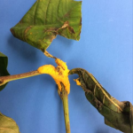
Oak wilt
Marginal leaf bronzing or tanning is often an early symptom of oak wilt.
I often think of oak wilt as the “Dutch elm disease of oaks” because there are many similarities between the two diseases. The oak wilt pathogen (the fungus Ceratocystis fagacearum) is often initially introduced into an area via insect (several types of sap beetles can be involved). These insects are attracted to wounded trees and, if they carry the oak wilt pathogen, can drop spores of the fungus off as they fed on sap oozing from wounds. Because of sap beetle transmission, I typically recommend pruning oaks only when they are dormant. In particular, the “January thaw” period in mid-winter is a good time to prune because it is warm enough so that you will not freeze to death while pruning, but not warm enough that the oak wilt fungus and sap beetles will be active. If for some reason you need to prune an oak during the growing season, you should IMMEDIATELY paint over pruning wounds to make them unattractive to sap beetles (which can visit wounds within 10 minutes of their formation). You can use a commercial pruning paint if you like, but latex paint (left over from painting the rooms of your house) will work as well.
Once the oak wilt fungus colonizes a tree, branches will begin to wilt and the tree will eventually die. In wooded areas, the oak wilt fungus can move from oak tree to oak tree via root grafts, causing major tree loss. Management of oak wilt in woodlots and forested areas typically involves establishing a perimeter around infected trees, trenching around this perimeter to sever root grafts and then removing oak trees within the trenched area. In landscape settings, single, healthy specimen oaks can be treated with fungicide injections (typically made every two years) to help prevent infection.
Making oak wilt management a challenge is the fact that some people diagnose oak wilt based on visual symptoms. I do not agree with this method of assessment, and personally will only diagnose oak wilt if I can grow the oak wilt fungus from symptomatic oak branches or trunk tissue, or detect Ceratocystis fagacearum DNA in this tissue. The danger of visual diagnosis is that there are other diseases and insect problems that can cause branch dieback symptoms that somewhat mimic those of oak wilt. In particular, I worry about misdiagnosis of Armillaria root disease as oak wilt, because trenching (advocated for oak wilt management) wounds roots and wounded roots are a primary entry point for Armillaria (the fungus that causes Armillaria root disease). Another problem that mimics oak wilt is damage due to two-lined chestnut borer, an insect pest that tends to attack oak trees that are under stress. Fungicide injections for oak wilt management are a waste of time, effort and money if the real problem is an insect pest such as two-lined chestnut borer. Proper diagnosis is the first step in developing a successful disease and insect management strategy for oak wilt (or any disease for that matter).
Verticillium wilt
Sudden yellowing, wilting and death of leaves and branches, particularly starting in one section of a tree or shrub, is a typical symptom of Verticillium wilt.
Verticillium wilt is disease that can affect a wide range of woody ornamentals including, but not limited to, maples (particularly Japanese and Korean maple), ash, redbud, magnolia, and smokebush/smoketree. The disease can also cause problems in vegetables (e.g., potato, tomato, pepper, vine crops and especially eggplant) as well as herbaceous ornamentals (I diagnosed Verticillium wilt in purple coneflower just recently). Verticillium (typically Verticillium dahliae) is soilborne and can be introduced into a location via contaminated soil, mulch (be cautious of using mulch composed of chipped up street trees that might have died from Verticillium wilt) or even leaves that have fallen from infected trees and been blown into an area. The fungus infects through roots, colonizing and blocking the xylem, resulting in branch dieback. In particularly susceptible trees (e.g., Japanese maples) and vegetables (e.g., eggplant), death can follow very rapidly. Proper diagnosis of Verticillium wilt is important because if Verticillium is present at a location, use of Verticillium immune or resistant plants is the best method to prevent problems in the future. That said, over the past three years, the PDDC has documented previously unreported hosts for Verticillium including seven-son flower, wafer ash, buttonbush and Eastern leatherwood.
On July 26, late blight (caused by the water mold Phytophthora infestans) was formally diagnosed in Wisconsin for the first time in 2017. The late blight sample was of infected tomato fruits from Waukesha County. Late blight attacks both potatoes and tomatoes, and unchecked the disease can rapidly kill plants. Late blight is the disease that caused the Irish potato famine in the 1840’s, resulting in the starvation of approximately 1 million Irish and the mass emigration of approximately another 1 million Irish, many to the US.
Nowadays, late blight can wipe out home garden tomatoes and potatoes, and can have a huge impact on fresh market tomato production. Most importantly in Wisconsin however is late blight’s potential impact on commercial potato production. Wisconsin is a leading potato producer in the US with greater than 60,000 acres in production in 2016.
Symptoms of late blight on potato and tomato leaves and stems typically appear as somewhat large, dark, oily areas, sometimes with a lighter border.
On tomato fruits, the disease often appears as large, leathery areas with somewhat wavy margins and sometimes visible concentric rings. The underside of infected leaves will typically have a fuzzy white-gray appearance, an indication that pathogen is sporulating. Sporulation also occurs on infected fruits, but can be more difficult to see. The disease progresses rapidly and kills plants. The pathogen can eventually also infect potato tubers where it can survive over the winter.
On tomato leaves (left), late blight leads to brown-black, water-soaked, oily areas that may have a white-gray fuzzy look. On tomato fruits (right), late blight leads to large, often sunken, golden- to chocolate-brown, firm spots with distinct rings.
Because of the importance of late blight to Wisconsin agriculture, the PDDC offers free diagnosis of suspect late blight potato and tomato samples. If you see anything that you think is late blight on potato or tomato (or even if you don’t have what you think is late blight on potato or tomato, but want to know what your potato or tomato problem is), send in a sample, invoke the words “late blight” and the diagnosis is free. To submit a sample, place symptomatic leaves, stems, fruits, and/or tubers in sealable plastic bags. DO NOT wrap the sample in wet paper toweling as this can accelerate the deterioration of the tissue and make diagnosis more difficult. Place the bagged sample(s) in a sturdy box with lots of padding and mail the sample to the:
PDDC
Department of Plant Pathology
University of Wisconsin-Madison
1630 Linden Drive
Madison, WI 53706-1598
Be sure to include complete contact information (i.e., complete mailing address, phone number and email address). If the sample is positive for late blight, PDDC staff will contact you via phone to provide guidance on how to manage the problem. All submitters will receive a written report outlining any disease problems in their samples. Samples positive for late blight will be forwarded to the lab of Dr. Amanda Gevens (the UW-Madison/Extension vegetable pathologist) for genotype testing. There are numerous variants (genotypes) of Phytophthora infestans and knowing which variant(s) [or genotype(s)] are present in Wisconsin can provide critical control information for commercial potato and tomato producers. Certain variants of Phytopthora infestans are resistant to certain fungicides; others are not.
To learn more about late blight and its management in home garden, check out the “Late Blight” University of Wisconsin Garden Facts in the fact sheets section of the PDDC website:
Also, be sure to follow the PDDC on Facebook and Twitter @UWPDDC for updates on plant diseases such as late blight, as well as to learn about PDDC educational events.
For additional resources on late blight (particularly for commercial growers) see the following links:
In my June web article, I mentioned cedar-apple rust and other Gymnosporangium rusts as diseases that I expected to see a lot of this year. This has certainly been the case over the last month. What has surprised me (and I probably shouldn’t be surprised given our continuing wet weather) is the plethora of other rust diseases that I have seen in the clinic this season. Here’s a rundown some of the more interesting examples of rust diseases that I’ve seen this year.
This is an alternating rust where the pathogen requires two different plants to complete its life cycle. In this case the hosts are white pine and Ribes species (in particular gooseberries and currants). The white pine phase of this disease reared its head in May in the Madison area (see my PDDC Facebook page post from May 20) and the Ribes phase of the disease should be in full force right now. Watch for orange, powdery masses of spores of the undersides of gooseberry and currant leaves. Management of this disease relies on not growing white pines and gooseberries/currants in close proximity.
Gymnosporangium rusts have definitely had a big year in 2017 and like white pine blister rust are alternating rusts. Check out apples, crabapples and hawthorns right now for characteristic yellow-orange leaf spots. I posted photos of the juniper stage of this disease on the PDDC Facebook page back on May 20. If our weather continues to be wet, I expect a banner year for the juniper stage of the disease next spring. Management of Gymnosporangium rusts is also most successful when the two hosts are not grown near one another.
Crown rust
Crown rust
This alternating rust has been particularly dramatic on buckthorn this year (see photos posted on the PDDC Facebook page post on June 22). There are several variants of the crown rust fungus and each variant has a specific grass alternate host. The most common alternate hosts that I encounter are oats (in agricultural settings) and turfgrass (in urban settings). If you need another reason to eradicate buckthorn (in addition to the fact that this plant is incredibly invasive), control of crown rust is that reason.
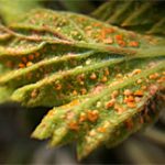
Orange rust
Orange rust
This rust requires just a single host (raspberry) to complete its life cycle, and leads to stunting of raspberry plants, yellowing of raspberry leaves, and eventual formation of masses of orange powdery spores on the undersides of leaves (see the PDDC Facebook page post on this disease from June 3 for photos). Unfortunately the orange rust fungus becomes systemic in plants (surviving as fungal hyphae/threads) and management requires plant removal and destruction.
Mayapple rust
Mayapple rust
This is another single host rust, with the fungus completing its life cycle on mayapple. Watch for angular (i.e., vein-bordered) yellow leaf spots with spores of the fungus produced directly beneath these spots on the undersides of the leaves. The resting spores produced by this fungus are two-celled and spiny (see the PDDC Facebook page post from June 30 for photos). This rust can be managed by a combination of removal of infected plants (as they go dormant for the season) and timely fungicide applications the following growing season as new plants emerge.
Bean rust
Bean rust
This is another single host (snap/pole beans) rust that I have seen in the past, but I have only ever seen the later stages of the disease where the fungus produces orange and brown-colored spores (called urediniospores and teliospores respectively). This year I got to see (for the first time ever) a third type of spore produced by the fungus (called aeciospores). These spores are white and are produced in rings of fruiting bodies (i.e., reproductive structures) on the underside of bean leaves (see the PDDC Facebook page post from June 30 for photos). This disease is relatively uncommon in home gardens, most likely because many snap bean varieties have at least some resistance to the disease.
To learn more about plant diseases and their management, explore the Plant Disease Diagnostics Clinic (PDDC) website (https://pddc.qa.webhosting.cals.wisc.edu/) or follow the PDDC on Facebook and Twitter @UWPDDC.
May has been a fairly wet month in many parts of Wisconsin. When spring rains overlap with leaf emergence on broad-leafed trees and shrubs, expect leaf diseases to run rampant over the summer. Some of diseases that I have already seen or I am expecting to see this year include:
Anthracnose refers to a large group of fungal leaf diseases. There are many different types of anthracnose fungi and they are somewhat host specific. However, all of these fungi tend to cause irregular, blotchy necrotic (i.e., dead) areas on leaves. If anthracnose occurs early (on leaves that are not fully expanded), leaves can become cupped and curled. On some trees (white oaks come to mind), anthracnose can be so severe that it will cause defoliation, but typically these trees will releaf and by July, you would never know the trees had anthracnose earlier in the year.
This is a disease I most commonly see on maples. The distinctive symptoms (either large, solid tar-like spots, or circular, diffuse clusters of smaller tarry spots) typically are not visible until later in the season. However, check maple leaves (particularly on Norway maples) right now for small yellow spots that are a clue that infections have already occurred. If you have a good (10X or 20X) hand lens, you may be able to see very small, tarry spots in the middle of the yellow areas. Continue to watch for more spectacular symptoms to develop as the summer progresses.
Leaf distortions and discoloration typical of peach leaf curl.
If you have a peach tree with curled, cupped and bubbly looking leaves, you have this disease. The distorted leaves often have a pinkish and/or yellowish color. There is nothing else that will cause these sorts of symptom on peach leaves.
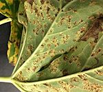
Leaf spots typical of apple scab on apple. Similar lesions occur on fruits.
I expect a banner year for this disease on apple and crabapple trees. The causal fungus survives in apple and crabapple leaf litter and releases spores during wet periods as leaves are beginning to emerge. Initial infections lead to dark, roughly circular leaf spots with somewhat feathery edges. Spores are produced in the infected areas leading to additional infections. Eventually entire leaves can look gray/black and sooty. Highly susceptible apple and crabapple varieties usually totally defoliate due to the disease by mid-season. Unfortunately the affected trees do not releaf.
Circular, yellow-orange diseased areas typical of cedar-apple rust on apple.
I received a number of photos of the juniper stage of this fungal disease earlier in the spring, and I posted several of these photos to my clinic Facebook page. The orange, marmalade-like masses that form on junipers produce spores that infect leaves of apples, crabapples and hawthorns. The leaf spots that eventually form (they are at their most vibrant in July and August) are often roughly nickel-sized and bright yellow, orange or maroon (on red-foliaged varieties of crabapples). Spores produced in these spots eventually reinfect junipers, thus completing the pathogen’s life cycle. Also watch for spiny, salmon-colored fruit on hawthorns, a variation of the disease called cedar-quince rust.
To learn more about these diseases (as well as other plant diseases) and their management, explore the Plant Disease Diagnostics Clinic (PDDC) website (https://pddc.qa.webhosting.cals.wisc.edu/) or follow the PDDC on Facebook and Twitter @UWPDDC.
If you are like many vegetable gardeners, you have transplants growing in your basement under artificial lights and are chomping at the bit to get those plants out into your garden.
Before you do that, here are a few things to think about to make your summer vegetable garden more successful.
Finish any last minute garden clean up.
If you have leftover vegetable plant debris in your garden, remove it now. These leftovers are where disease-causing fungi and bacteria overwinter and they can serve as a source of pathogens that can infect your new garden plants. Burn (where allowed), deep bury or hot compost these materials.
Clean other gardening items.
Disease-causing organisms can survive the winter on gardening tools, stakes and cages. These items should be decontaminated before using them again:
Remove any clinging soil or plant debris.
Use 70% alcohol (e.g., rubbing alcohol, spray disinfectants) or 10% bleach to complete the process:
Treat metal items with alcohol, either dipping them for 30 seconds or spraying them until they drip and allowing them to air dry.
For non-metal items, soak in bleach for 20-30 minutes, then rinse thoroughly to remove bleach residues. Tip: Be sure to wear old clothes, rubber gloves and eye protection when working with bleach.
Invest in a soaker or drip hose.
If you use a sprinkler to water, you are getting leaves wet and this provides an environment that is perfect to get diseases started. Soaker and drip hoses keep water off of leaves and apply it into the soil where it is most useful.
Map out your garden.
One way to reduce disease problems is to make sure you move vegetables around in your garden each year. This is called rotation and helps prevent buildup of disease-causing organisms in the soil.
For details on how to use rotation most effectively, check out the University of Wisconsin Garden Facts fact sheet “Using Crop Rotation in the Home Vegetable Garden.” Each year make a map of where you have specific vegetables and keep these maps so that you know where to rotate your vegetables each year.
Keep a journal.
Write down observations of what goes on in your garden and when.
When did you plant?
When did seedlings emerge?
When did plants bloom?
When did they set fruit? When did you harvest?
Did you see particular diseases or insect pests?
When did they start?
All of this sort of information can be helpful in planning your garden in the future. After several years, you will also get a sense of what disease and insect problems are common and when they typically arrive. Armed with this information, you can more efficiently and effectively develop management strategies.
Enjoy growing the old standards that you love, but also do not be afraid to try new (and what may seem like exotic) vegetables. Trying new things keeps gardening fresh and exciting, and exposes you to new flavors and cuisines.
Have fun!
That’s what gardening should be all about.
Additional Resources
To learn more about plant diseases and their management, explore the Plant Disease Diagnostics Clinic (PDDC) website (https://pddc.qa.webhosting.cals.wisc.edu/) or follow the PDDC on Facebook and Twitter @UWPDDC.
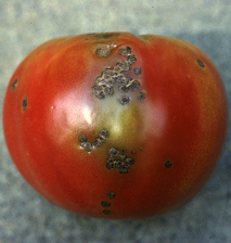
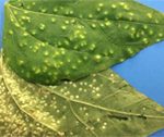
Sunken, scabby bacterial spot lesions on ripening tomato fruit. (Photo courtesy of Mary Ann Hansen, Virginia Polytechnic Institute and State University)
What is bacterial spot?
Bacterial spot of tomato is a potentially devastating disease that, in severe cases, can lead to unmarketable fruit and even plant death. Bacterial spot can occur wherever tomatoes are grown, but is found most frequently in warm, wet climates, as well as in greenhouses. The disease is often an issue in Wisconsin.
What does bacterial spot look like?
Bacterial spot can affect all above ground parts of a tomato plant, including the leaves, stems, and fruit. Bacterial spot appears on leaves as small (less than ⅛ inch), sometimes water-soaked (i.e., wet-looking) circular areas. Spots may initially be yellow-green but darken to brownish-red as they age. When the disease is severe, extensive leaf yellowing and leaf loss can also occur. On green fruit, spots are typically small, raised and blister-like, and may have a yellowish halo. As fruit mature, the spots enlarge (reaching a maximum size of ¼ inch) and turn brown, scabby and rough. Mature spots may be raised or sunken with raised edges. Bacterial spot symptoms can be easily confused with symptoms of another tomato disease called bacterial speck. For more information on this disease, see UW Plant Disease Facts D0011, Bacterial Speck of Tomato.
Where does bacterial spot come from?
Bacterial spot of tomato is caused by Xanthomonas vesicatoria, Xanthomonas euvesicatoria, Xanthomonas gardneri, and Xanthomonas perforans. These bacterial pathogens can be introduced into a garden on contaminated seed and transplants, which may or may not show symptoms. The pathogens enter plants through natural openings (e.g., stomates), as well as through wounds. Disease development is favored by warm (75° to 86°F), wet weather. Wind-driven rain can contribute to more severe disease as the pathogens are splashed and spread to healthy leaves and fruit. Bacterial spot pathogens can survive well in tomato debris, but they survive very poorly in soil when not associated with debris.
On tomato leaves, bacterial spot leads to small, angular (i.e., straight-edged) spots with yellow haloes. (Photo courtesy of Michelle Grabowski, University of Minnesota Extension)
How do I save plants with bacterial spot?
A plant with bacterial spot cannot be cured. Remove symptomatic plants from your garden or greenhouse to prevent the spread of bacteria to healthy plants. Burn (where allowed by local ordinance), bury or hot compost the affected plants, and DO NOT eat symptomatic fruit. Although bacterial spot pathogens are not human pathogens, the fruit blemishes that they cause can provide entry points for human pathogens that could cause illness.
How can I prevent bacterial spot in the future?
Plant pathogen-free seed or transplants to prevent the introduction of bacterial spot pathogens on contaminated seed or seedlings. If a clean seed source is not available or you suspect that your seed is contaminated, consider treating the seed in hot water prior to planting to eliminate the pathogens. For details on this process, including the proper temperature and length of treatment, see UW Plant Disease Facts D0064, Hot-Water Seed Treatment for Disease Management. To keep leaves dry and to prevent the spread of the pathogens, avoid overhead watering (e.g., with a wand or sprinkler) of established plants. Instead use a drip-tape or soaker-hose. Also, to prevent spread, DO NOT handle plants when they are wet (e.g., from dew), and routinely sterilize tools with either 10% bleach solution or (better due to its less corrosive properties) 70% alcohol (e.g., rubbing alcohol). Where bacterial spot has been a recurring problem, consider using preventative applications of copper-based products registered for use on tomato, especially during warm, wet periods. Keep in mind however, that if used excessively or for prolonged periods, copper may no longer control the disease. Be sure to read and follow all label instructions of the product that you select to ensure that you use it in the safest and most effective manner possible. Burn (where allowed by local ordinance), bury or hot compost tomato debris at the end of the season. Wait at least one year before planting tomatoes in a given location again, and remove and burn, bury or hot compost any volunteer tomatoes that come up in your garden.
For more information on bacterial spot of tomato:
Contact the University of Wisconsin Plant Disease Diagnostics Clinic (PDDC) at (608) 262-2863 or pddc@wisc.edu.
An EEO/Affirmative Action employer, University of Wisconsin-Madison Division of Extension provides equal opportunities in employment and programming, including Title IX and ADA requirements. This document can be provided in an alternative format by calling Brian Hudelson at (608) 262-2863 (711 for Wisconsin Relay).
References to pesticide products in this publication are for your convenience and are not an endorsement or criticism of one product over similar products. You are responsible for using pesticides according to the manufacturer’s current label directions. Follow directions exactly to protect the environment and people from pesticide exposure. Failure to do so violates the law.
Thanks to Amanda Gevens, Gary Marks, Jessamyn Perlus, Carol Shirk, Bryan Webster and Ken Williams for reviewing this document.
A complete inventory of UW Plant Disease Facts is available at the University of Wisconsin-Madison Plant Disease Diagnostics Clinic website: https://pddc.qa.webhosting.cals.wisc.edu.

 Given that turkey is an animal, rather than a plant-based food, one would think nothing could go wrong. Ah, but then we have to consider the stuffing. Stuffing is made, in part, from cereal grains (e.g., wheat) and one disease that could cause issues is
  I have never met a potato product that I didn’t like, but mashed potatoes are my favorite. I think this is because at Thanksgiving, my family’s tradition is to serve them not with gravy, but with homemade egg noodles cooked in turkey/chicken broth. Everyone thinks this tradition is weird at best, but once folks have experienced “noodle gravy” they become converts. From a plant pathology standpoint, the most famous disease that might limit my access to potatoes is
  I have a great recipe for cranberry/butter/brown sugar glazed carrots I like to serve at Thanksgiving (the recipe is at the bottom of this page). But if I store carrots too long in my refrigerator, they can turn to a slimy mess and be totally unusable for this delicious recipe. The disease that causes this degradation is
  What Thanksgiving dinner would be complete without a cranberry dish of some kind? Cranberry relish, cranberry salad and cranberry bread are all delicious additions to a Thanksgiving meal. But cranberries can have their disease problems as well. Wisconsin is the largest cranberry producing state in the US, so I see a fair share of cranberry samples come into my clinic. The most common problems I see are a variety of
  Pumpkin pie is another of my favorites and I can scarf an entire pie in one sitting. So, any disease of pumpkins is on my hit list. The most common disease I encounter on pumpkins is
  One of the easiest and most effective ways to help manage plant diseases is good fall cleanup of your yard and garden. Many common fungal and bacterial plant pathogens, particularly those that cause leaf diseases, survive Wisconsin winters in leaf litter from trees and shrubs, as well as on herbaceous plant parts that have died back for the winter. Disease-causing organisms can also survive on common gardening items like pots, stakes and tools. So, as the temperatures cool and plants begin to go dormant for the season, here are a couple of things to think about doing to put your gardens and landscape to bed for the winter and have a head start on your gardening for next year.
 The safest way to dispose of these materials is typically to take them to a local yard waste center (if there is one available in your community) where they can be properly composted. If not, other options for disposing of this material include burning (not particularly environmentally friendly, but an option if allowed by local ordinance), burying (make sure the material is completely covered by approximately two inches of soil) or hot
  For clay or ceramic pots, wash the pots well with soapy water (to remove any remaining soil), then soak them for 30 minutes in a 10% bleach solution. Rinse your pots well to remove bleach residues, allow them to air dry, then store them in a clean location where they will not be recontaminated. Decontamination of plastic pots (or other plastic items like stakes) is often more challenging. Often bleaching will not be effective and your best option may be to throw out plastic items and buy new ones next year. Decontaminate gardening tools by dipping them for a minimum of 30 seconds in 70% alcohol (e.g., rubbing alcohol) or spraying them until they drip with a spray disinfectant that contains approximately 70% alcohol. As with decontaminated pots, store clean tools in a location where they will not be recontaminated.


 If you are like many vegetable gardeners, you have transplants growing in your basement under artificial lights and are chomping at the bit to get those plants out into your garden.